
Clinical Study
Proof-of-Principle Study
Discover how innovative digital photography and sclera color analysis formed the basis of our app. Our proof-of-principle study at UCLH demonstrated the potential of this non-invasive, accessible technique to improve early detection and treatment outcomes for newborns.
Study Overview
Our clinical study at University College London Hospitals (UCLH) in 2015 aimed to validate the use of digital photography and sclera color analysis for screening neonatal jaundice. Conducted by researchers from UCL’s Department of Medical Physics & Biomedical Engineering and the Neonatal Care Unit at UCLH, the study involved 133 newborns. We found that digital photography performed well comparitively against traditional methods like TCBs. This shows us that this method could become a low-cost, accessible tool for early jaundice detection, especially in low-resource settings.
Methodology
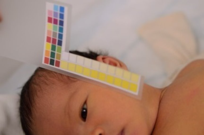
- Image Capture: Photographs of newborns’ eyes were taken using a Nikon D3200 camera with a 60 mm macro lens. Manual focus and auto exposure settings were used and the ISO was set to 1600. White balance was maintained for consistency.
- Calibration: A customised color chart was used to ensure accurate color readings. Images were captured in RAW format to preserve tonal information.
- Subjects: Of the 133 newborns photographed, 110 samples were used for analysis due to consistent lighting conditions. The sample included 67 white and 43 non-white infants.
Results
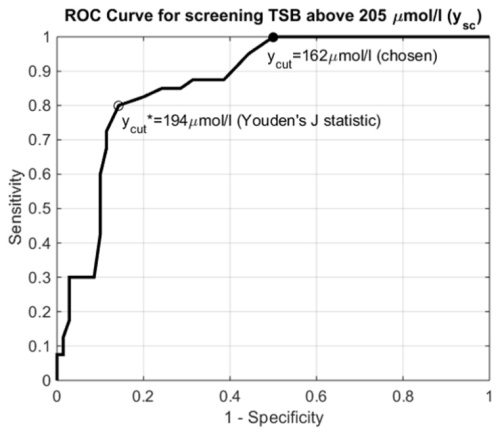
We found that using sclera color to predict TSB levels provided reasonably high correlations (r = 0.75, ρ = 0.72). The ROC curve analysis showed an area under the curve (AUC) of 0.87, indicating the method’s potential as a screening tool.
Discussion
- Ambient Lighting: We found that ambient lighting significantly affects prediction accuracy. Using images taken in controlled lighting conditions improved correlation results.
- Comparisons: We compared our results with those from other techniques like transcutaneous bilirubinometers (TcB), showing that while correlations were slightly lower, the method’s non-contact nature and potential for smartphone integration offer significant advantages.
Conclusion
This study demonstrated that sclera color analysis using digital photography is a promising technique for neonatal jaundice screening. With further improvements in ambient lighting control and algorithm refinement, this method could become a low-cost, accessible tool for early jaundice detection, especially in low-resource settings.
Related Publications
November 2015 – Screening neonatal jaundice based on the sclera color of the eye using digital photography
January 2019 – Jaundice Eye Color Index (JECI): quantifying the yellowness of the sclera in jaundiced neonates with digital photography